Procedure of Intravascular Lithotripsy
The procedure of Intravascular Lithotripsy (IVL) involves several steps:
Patient Preparation:
The patient is prepared for the procedure, which typically involves obtaining consent, reviewing medical history, and administering any necessary medications.
Access and Guidewire Placement:
A local anesthetic is administered, and a vascular access site, often the femoral or radial artery, is prepared. A specialized IVL catheter is then advanced over a guidewire to the target artery under fluoroscopic guidance.
Lesion Assessment:
Angiography is performed to visualize the calcified lesion within the artery. Intravascular imaging techniques such as intravascular ultrasound (IVUS) or optical coherence tomography (OCT) may also be used to assess lesion morphology and guide treatment planning.
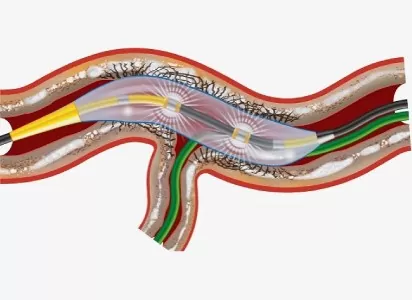
Balloon Positioning:
Once the calcified lesion is identified, the IVL catheter is positioned proximal to the lesion site. The IVL catheter consists of a balloon with integrated lithotripsy emitters.
Lithotripsy Activation:
The IVL system is activated, delivering sonic pressure waves through the balloon emitters to the calcified plaque. These waves disrupt the calcium deposits, creating micro-fractures within the plaque while minimizing trauma to the surrounding vessel wall.
Balloon Dilatation (Optional):
Following lithotripsy treatment, balloon angioplasty may be performed to further dilate the artery and optimize lumen diameter. This step helps ensure adequate stent delivery and expansion.
Stent Placement:
If indicated, a stent is deployed to scaffold the treated segment of the artery and restore blood flow. The stent is positioned across the lesion site and expanded using balloon inflation.
Final Assessment:
Angiography is repeated to assess the results of the IVL treatment and confirm optimal stent deployment. Any residual stenosis or dissections are addressed as needed.
Closure and Post-procedural Care:
Once the procedure is complete, the IVL catheter is removed, and hemostasis is achieved at the vascular access site. The patient is monitored for a period to detect any complications and may be prescribed medications to prevent thrombosis or manage underlying cardiovascular conditions.